Ultra-High-Frequency ECG in Cardiac Pacing and Cardiac Resynchronization Therapy: From Technical Concept to Clinical Application
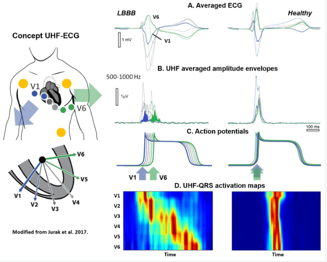
"UHF-ECG can determine a tailored resynchronization approach for the optimization of CRT and holds promise beyond CRT for the risk stratification of ventricular arrhythmias." by Uyên Châu Nguyên et al.

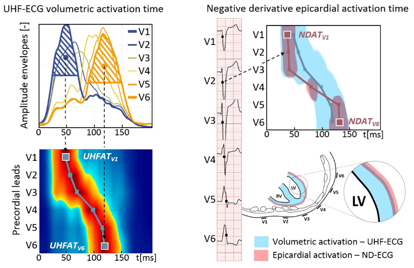
Ultra-high-frequency ECG volumetric and negative derivative epicardial ventricular electrical activation pattern
"Combining new UHF-ECG with conventional ND-ECG offers a more comprehensive analysis of heart activation throughout the muscle wall compared to using just one method." by Pavel Leinveber et al.

Non-invasive Conduction System Pacing guidance
Regular 12(14)-lead ECG with real-time visualization
It could not be easier ...