How UHF-ECG works
Real-time, non-invasive, regular 12(14) lead ECG setup
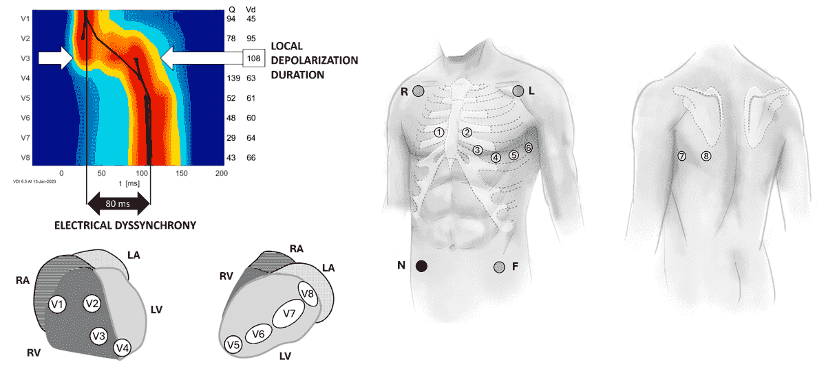
12(14) lead ECG mapping
Standard 12(14) lead ECG setup is all to start to measure.
Redcolor indicates the time at which the greatest number of myocardial cells under
the electrode depolarize.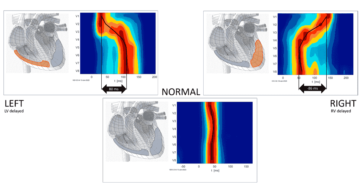
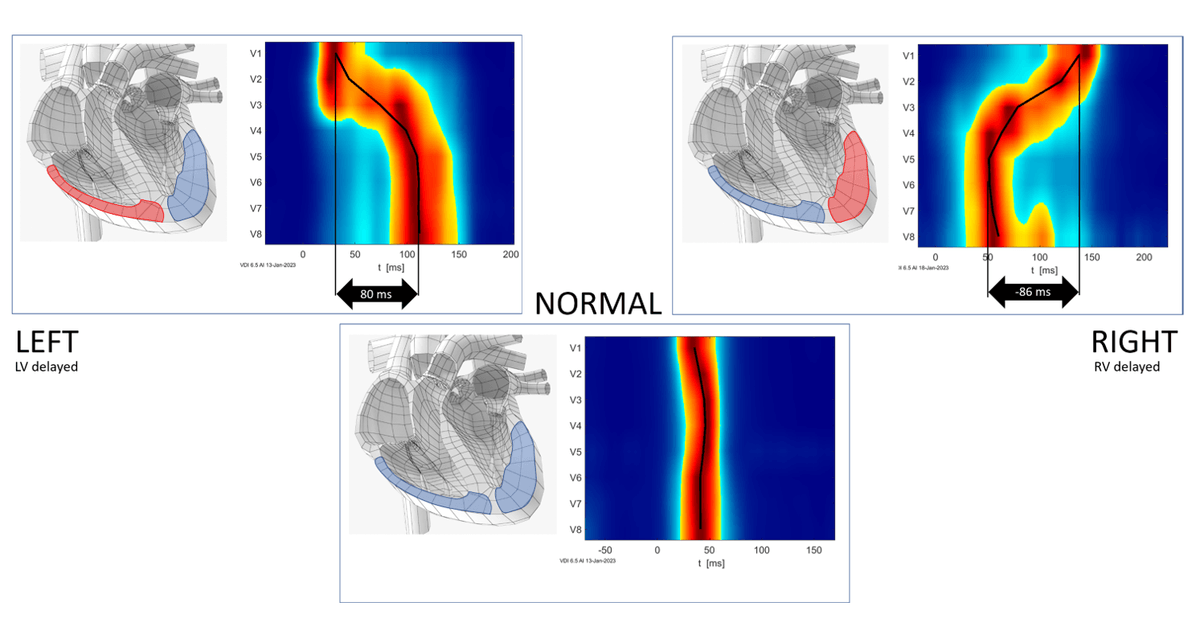
Activation patterns
Activation maps clearly show the ventricular activation pattern.
VDI UHF-ECG Atlas
Synchronous UHF-ECG activation example
All ventricular segments under chest leads are activated almost at the same time.
Dyssynchronous UHF-ECG activation
Dyssynchronous activation shows a significant time delay between individual chest leads.
UHF-ECG FAQs
How is UHF-ECG different from a standard ECG?
It's completely different and not a typical P-QRS-T ECG. It captures frequencies up to 1000 Hz during QRS, providing information about ventricular depolarization in selected ventricular segments that a standard ECG cannot detect.
What does UHF‑ECG show?
It provides an activation map that visualizes the timing and sequence of ventricular activation. The main numerical parameters include electrical dyssynchrony and local activation duration.
Is UHF‑ECG safe?
Yes. It is completely passive and non‑invasive, using standard chest electrodes.
When is UHF‑ECG useful?
It supports CRT, conduction system pacing, assessment of dyssynchrony, and
evaluation of conduction abnormalities.UHF-ECG Bibliography
Main Publications
Curila, K., Mizner, J., Morava, J., Smisek, R., Vesela, J., Sussenbek, O., Stros, P., Kupec, J., Waldauf, P., Leinveber, P.,Poviser, L., Nagy, L., Cerny, J., Bitmanova, B., Jurak, P., & Polasek, R. (2025). Prospective randomized trial of conduction system pacing vs right ventricular pacing for patients with atrioventricular block; PragueCSP trial. Heart Rhythm. https://doi.org/10.1016/j.hrthm.2025.05.036
Mizner,J., Beela, A., Linkova, H., Vesela, J., Sussenbek, O., Stros, P., Smisek, R., Jurak, P., Leinveber, P., Lipoldova, J., Nagy, A., Waldauf, P., Lumens, J.,Vernooy, K., Prinzen, F., & Curila, K. (2025). Electrical and mechanicalinterventricular dyssynchrony coupling in patients with bradycardia: A UHF-ECG validation trial. HeartRhythm. https://doi.org/10.1016/j.hrthm.2025.02.031
Moraleda-Salas,M. T., Amigo-Otero, E., Esteve-Ruiz, I., Arce-León, Á., Carreño-Lineros, J. M., Izaga Torralba, E., Navarro Roldan, F., & Moriña-Vázquez, P. (2024). Early improvement in cardiac functionand dyssynchrony after physiological upgrading in pacing-induced cardiomyopathy. Pacingand Clinical Electrophysiology. https://doi.org/10.1111/pace.15126
Verstappen, A. A. A., Hautvast, R., Jurak, P., Bracke, F. A., & Rademakers, L. M. (2024). Ventricular dyssynchrony imaging, echocardiographic and clinical outcomes of left bundle branch pacing and biventricular pacing. Indian Pacing and Electrophysiology Journal, 24(3), 140-146. https://doi.org/10.1016/j.ipej.2024.04.007
Curila, K, Poviser, L, Stros, P. et al. LVSP and LBBP Result in Similar or Improved LV Synchrony and Hemodynamics Compared to BVP. J Am Coll Cardiol EP. 2024 Jul, 10 (7_Part_2) 1722–1732.
https://www.jacc.org/action/showCitFormats?doi=10.1016/j.jacep.2024.04.022
Nguyên U.C, Rijks J.H.J, Plesinger F, et al. Ultra-High-Frequency ECG in Cardiac Pacing and Cardiac Resynchronization Therapy: From Technical Concept to Clinical Application. J. Cardiovasc. Dev. Dis. 2024, 11(3), 76. doi.org/10.3390/jcdd11030076.https://www.mdpi.com/2308-3425/11/3/76
Curila, K., Jurak, P., & Varma, N. (2023). Resynchronization for shifting conduction patterns - When a coronary sinus lead is not enough. Indian Pacing and Electrophysiology Journal, 23(6), 214-215. https://doi.org/10.1016/j.ipej.2023.08.005
Sussenbek, O., Rademakers, L., Waldauf, P., Jurak, P., Smisek, R., Stros, P., Poviser, L., Vesela, J., Plesinger, F., Halamek, J., Leinveber, P., Herman, D., Osmancik, P., & Curila, K. (2023). Left bundle branch area pacing results in more physiological ventricular activation than biventricular pacing in patients with left bundle branch block heart failure. European Heart Journal Supplements, 25(Suppl E), E17-E24. https://doi.org/10.1093/eurheartjsupp/suad109
Leinveber P, Halamek J, Curila K et al. Ultra-high-frequency ECG volumetric and negative derivative epicardial ventricular electrical activation pattern. Sci Rep 14, 5681 (2024). doi.org/10.1038/s41598-024-55789-w
https://www.nature.com/articles/s41598-024-55789-wCurila K, Jurak P, Prinzen F.,et al. Bipolar anodal septal pacing with direct LBB capture preserves physiological Ventricular activation better than unipolar left bundle branch pacing. Front.Cardiovasc. Med.,22 March 2023.
https://www.frontiersin.org/articles/10.3389/fcvm.2023.1140988/full
Mizner J, Jurak P, Linkova H, Smisek R, Curila K. Ventricular dyssynchrony and pacing-induced cardiomyopathy in patients with pacemakers, the utility of ultra-high-frequency ECG and other dyssynchrony assessment tools.Arrhythmia &Electrophysiology Review.2022;11. doi:10.15420/aer. 2022. 01https://www.ncbi.nlm.nih.gov/pmc/articles/PMC9376832/
Jurak P, Bear L, Nguyên U, etal. 3-DimensionalVentricular Electrical Activation Pattern Assessed from A Novel High-Frequency Electrocardiographic Imaging Technique: Principles and Clinical Importance. Scientific Reports. 11. 2021https://www.nature.com/articles/s41598-021-90963-4
Curila K, Jurak P, JastrzebskiM, et al. Left bundle branch pacing compared to left ventricular septal myocardial pacing increases interventricular dyssynchrony but accelerates left ventricular lateral wall depolarization. Heart Rhythm.2021;18(8):1281-1289. doi:10.1016/j.hrthm.2021.04.025https://www.heartrhythmjournal.com/article/S1547-5271(21)00402-1/pdf
Curila K, Jurak P, Halamek J,et al. Ventricular Activation Pattern Assessment during Right Ventricular Pacing; Ultra-High-Frequency ECG Study. Journal of Cardiovascular Electrophysiology. 2021. https://onlinelibrary.wiley.com/doi/full/10.1111/jce.14985
Curila K, Prochazkova R, JurakP, et al. Both selective and nonselective His bundle, but not myocardial, pacing preserve ventricular electrical synchrony assessed by ultra-high-frequency ECG.Heart Rhythm.2020;17(4):607-614. doi:10.1016/j.hrthm.2019.11.016https://www.heartrhythmjournal.com/article/S1547-5271(19)31028-8/pdf
Jurak P, Curila K, LeinveberP, et al. Novel ultra‐high‐frequency electrocardiogram tool for the description of the ventricular depolarization pattern before and during cardiac resynchronization.Journal of Cardiovascular Electrophysiology.2019;31(1):300-307. doi:10.1111/jce.14299https://onlinelibrary.wiley.com/doi/full/10.1111/jce.14299
Plesinger F, Jurak P, HalamekJ, et al. Ventricular Electrical Delay Measured From Body Surface ECGs Is Associated With Cardiac Resynchronization Therapy Response in Left Bundle Branch Block Patients From the MADIT-CRT Trial (Multicenter Automatic Defibrillator Implantation-Cardiac Resynchronization Therapy).Circulation:Arrhythmia and Electrophysiology.2018;11(5). doi:10.1161/circep.117.005719https://www.ahajournals.org/doi/full/10.1161/CIRCEP.117.005719
Jurak P, Halamek J, Meluzin J,et al. Ventricular dyssynchrony assessment using ultra-high frequency ECG technique.Journal of Interventional Cardiac Electrophysiology.2017;49(3):245-254. doi:10.1007/s10840-017-0268-0 https://link.springer.com/article/10.1007/s10840-017-0268-0Other Publications
Prinzen FW, Jurak P, Leinveber P, Plesinger F, Curila K, Halamek J. Comparison of UHF-ECG with other noninvasive electrophysiological mapping tools for assessing ventricular dyssynchrony. In: 2021Computing in Cardiology (CinC).IEEE; 2021. http://dx.doi.org/10.23919/cinc53138.2021.9662706
Curila K, Jurak P, Leinveber P, et al. Physiological versus non-physiological cardiac pacing as assessed by Ultra-high-frequency electrocardiography. In: 2021Computing in Cardiology (CinC). IEEE; 2021. http://dx.doi.org/10.23919/cinc53138.2021.9662912
Plesinger F, Viscor I, Vondra V, et al. VDI Vision - Analysis of Ventricular Electrical Dyssynchrony in Real-Time. In: 2021Computing in Cardiology (CinC). IEEE; 2021. http://dx.doi.org/10.23919/cinc53138.2021.9662916
Koscova Z, Ivora A, Nejedly P, et al. QRS Complex Detection in Paced and Spontaneous Ultra-High-Frequency ECG. In: 2021Computing in Cardiology (CinC). IEEE; 2021. http://dx.doi.org/10.23919/cinc53138.2021.9662647
Jurak P, Leinveber P, Plesinger F, et al. Ultra-High-Frequency Electrocardiography. In: 2021 Computing in Cardiology (CinC).IEEE; 2021. http://dx.doi.org/10.23919/cinc53138.2021.9662795
Leinveber P, Halamek J, Jurak P, et al. The Ultra-High-Frequency QRS Dyssynchrony in the Assessment of Cardiac Resynchronization Therapy Effect. In: 2019 Computing in Cardiology Conference (CinC).2019. http://dx.doi.org/10.22489/cinc.2019.368
Matejkova M, Lipoldova J, Leinveber P, et al. Optimized CRT Stimulation Based on Ultra-High-Frequency QRS Analysis. In: 2019 Computing in Cardiology Conference (CinC). 2019. http://dx.doi.org/10.22489/cinc.2019.071
Halamek J, Leinveber P, Viscor I, et al. Cardiac Resynchronization Guided by Ultra-High-Frequency ECG Maps. In: 2019 Computing in Cardiology Conference (CinC). 2019. http://dx.doi.org/10.22489/cinc.2019.246
Halamek J, Leinveber P, Viscor I, et al. The relationship between ECG predictors of cardiac resynchronization therapy benefit. PLOS ONE.2019;14(5):e0217097. doi:10.1371/journal.pone.0217097
Halamek J, Leinveber P, Malik M, et al. High-Frequency QRS Analysis From Orthogonal Leads. In: 2018 Computing in Cardiology (CinC). 2018. http://dx.doi.org/10.22489/cinc.2018.051
Andrla P, Leinveber P, Châu Nguyên U, et al. Body-Surface Mapping Using High-Frequency ECG to Characterize Electrical Activation Delay. In: 2018 Computing in Cardiology. 2018. http://dx.doi.org/10.22489/cinc.2018.105
Jurak P, Châu Nguyên U,Viscor I, et al. Epicardial Isochrones from a New High-Frequency ECG Imaging Technique. In: 2018 Computing in Cardiology (CinC). 2018. http://dx.doi.org/10.22489/cinc.2018.088
Plesinger F, Jurak P, Halamek J, et al. veber P, Viscor I, Jurak P. A Method for Removing Pacing Artifacts From Ultra-High-Frequency Electrocardiograms. In: 2018 Computing in Cardiology (CinC).2018. http://dx.doi.org/10.22489/cinc.2018.106p://dx.doi.org/10.22489/cinc.2018.339
Andrla P, Plesinger F, Halamek J, Leinveber P, Viscor I, Jurak P. A Method for Removing Pacing Artifacts From Ultra-High-Frequency Electrocardiograms. In: 2018 Computing in Cardiology (CinC).2018. http://dx.doi.org/10.22489/cinc.2018.106
Halamek J, Leinveber P, Plesinger F, Matejkova M, Jurak P. Attenuation of QRS Power in the Frequency Range from 0.05 to 1 kHz. In: Computing in Cardiology (CinC). 2017. http://dx.doi.org/10.22489/cinc.2017.096-110
Plesinger F, Jurak P, Halamek J, et al. The VED Meter - a New Tool to Measure the Ventricular Conduction Abnormalities in Heart Failure Patients. In: Computingin Cardiology (CinC). 2017. http://dx.doi.org/10.22489/cinc.2017.377-059
Leinveber P, Halamek J, Jurak P. Ambulatory monitoring of myocardial ischemia in the 21st century—an opportunity for high-frequency QRS analysis. Journal of Electrocardiology. 2016;49(6):902-906. doi:10.1016/j.jelectrocard.2016.07.034
Jurak P, Leinveber P, Halamek J, et al. Biventricular Pacing Optimization by Means of the Dyssynchrony Parameter. In: 2016Computing in Cardiology (CinC).2016. http://dx.doi.org/10.22489/cinc.2016.051-263
Reichlova T, Jurak P, Halamek J, et al. Cardiac resynchronization efficiency estimation by new ultra-high-frequency ECG dyssynchrony descriptor. In: 2015 Computing in Cardiology (CinC).IEEE; 2015. http://dx.doi.org/10.1109/cic.2015.7410964
Jurak P, Halamek J, Plesinger F, et al. An additional marker of ventricular dyssynchrony. In: 2015 Computing in Cardiology (CinC). IEEE; 2015. http://dx.doi.org/10.1109/cic.2015.7408590
Plesinger F, Jurco J, Halamek J, Leinveber P, Reichlova T, Jurak P. Multichannel QRS Morphology Clustering - Data Preprocessing for Ultra-High-Frequency ECG Analysis. In: Proceedingsof the 3rd International Congress on Cardiovascular Technologies.SCITEPRESS - Science and and Technology Publications; 2015. http://dx.doi.org/10.5220/0005604200110019
Jurak P, Halamek J, Leinveber P, et al. Time-frequency interpretation of ultra-high-frequency QRS components. In: 20148th Conference of the European Study Group on Cardiovascular Oscillations (ESGCO).IEEE; 2014. http://dx.doi.org/10.1109/esgco.2014.6847526
Jurak P, Halamek J, Leinveber P, Vondra V, Soukup L, Vesely P, Sumbera J, Zeman K, Martinakova L, Jurakova T, Novak M. Ultra-high-frequency ECG measurement. In:2013, Computing in Cardiology (CinC).IEEE; 2013 http://cinc.mit.edu/archives/2013/pdf/0783.pdf